Question # 4
Which sonographic finding is most consistent with scrotal inflammation?
A.
Abscess
B.
Hydrocele
C.
Granuloma
D.
Hyperemia
Full Access
Answer:
D
Explanation:
Scrotal inflammation, such as epididymitis or orchitis, typically presents with increased blood flow (hyperemia) on color Doppler sonography. This finding reflects the inflammatory process and vascular dilation. Abscesses, granulomas, or hydroceles may be present but are not as consistent or specific for inflammation.
According to AIUM Practice Parameters and Rumack’s Diagnostic Ultrasound:
“In acute inflammation, color Doppler ultrasound demonstrates prominent hyperemia of the epididymis or testis.â€
[Reference:, Rumack CM, Wilson SR, Charboneau JW, Levine D. Diagnostic Ultrasound. 5th ed. Elsevier, 2017., AIUM Practice Parameter for Scrotal Ultrasound, 2020., —]
Question # 5
The absence of which sonographic finding indicates the acute process depicted in these images?
A.
Free fluid
B.
Ductal dilatation
C.
Hepatic vein thrombosis
D.
Cavernous transformation
Full Access
Answer:
D
Explanation:
The sonographic images depict an acute thrombotic process involving the portal venous system. The absence of cavernous transformation in the setting of portal vein thrombus indicates that the process is acute. In chronic portal vein thrombosis, collateral vessels form in the porta hepatis to bypass the obstruction, a process known as cavernous transformation.
Sonographic features suggesting acute portal vein thrombosis:
Echogenic thrombus within the portal vein lumen
Absence of flow on color Doppler
Enlarged portal vein diameter early in the process
No evidence of cavernous transformation (i.e., no serpiginous collateral vessels at porta hepatis)
Cavernous transformation is a hallmark of chronic portal vein thrombosis and takes weeks to months to develop. Therefore, its absence on ultrasound supports an acute diagnosis.
Differentiation from other options:
A. Free fluid: Non-specific and may or may not be present in hepatic vascular thrombosis.
B. Ductal dilatation: Related to biliary obstruction, not portal or hepatic venous thrombosis.
C. Hepatic vein thrombosis: Seen in Budd-Chiari syndrome, which affects hepatic outflow, not portal inflow.
[References:, Rumack CM, Wilson SR, Charboneau JW, Levine D. Diagnostic Ultrasound. 5th Edition. Elsevier, 2018. Chapter: Portal Venous System, pp. 105–108., American Institute of Ultrasound in Medicine (AIUM) Practice Parameter for the Performance of Hepatic Doppler Ultrasound Examinations, 2020., Radiopaedia.org. Cavernous transformation of the portal vein: https://radiopaedia.org/articles/cavernous-transformation-of-the-portal-vein, ]
Question # 6
Which sonographic finding indicates the need for immediate surgical intervention following testicular trauma?
A.
Intratesticular hematoma
B.
Discontinuity of the tunica albuginea
C.
Heterogeneity of the testicular parenchyma
D.
Increased testicular vascularity
Full Access
Answer:
B
Explanation:
The tunica albuginea is a dense fibrous capsule surrounding the testis. Discontinuity of the tunica albuginea on ultrasound is diagnostic of testicular rupture — a urologic emergency that requires immediate surgical repair to preserve testicular function and viability. Early surgical intervention within 72 hours has a high success rate for testicular salvage (up to 90%).
Intratesticular hematoma (A) may be managed conservatively if the tunica albuginea is intact.
Heterogeneity of the parenchyma (C) indicates injury but not necessarily rupture.
Increased vascularity (D) may be seen with inflammation or reperfusion but does not mandate surgery unless rupture is present.
Reference Extracts:
Dogra VS, Bhatt S. "Acute painful scrotum: ultrasound evaluation." Radiologic Clinics of North America. 2004; 42(2):349-363.
Middleton WD, Kurtz AB, Hertzberg BS.Ultrasound: The Requisites. 3rd ed. Elsevier, 2015.
—
Question # 7
Which condition is a common cause of biliary duct obstruction?
A.
Tumor
B.
Cholecystitis
C.
Pneumobilia
D.
Hepatitis
Full Access
Answer:
A
Explanation:
A tumor (such as cholangiocarcinoma, pancreatic head carcinoma, or metastases) is a common cause of biliary duct obstruction. It can compress or invade the bile ducts, leading to intrahepatic and extrahepatic duct dilatation.
Cholecystitis (B) typically affects the gallbladder but may rarely cause duct obstruction if complicated.
Pneumobilia (C) refers to air in the biliary tree, not obstruction.
Hepatitis (D) causes liver inflammation but not mechanical biliary obstruction.
Reference Extracts:
Rumack CM, Wilson SR, Charboneau JW, Levine D. Diagnostic Ultrasound. 5th ed. Elsevier, 2017.
Gore RM, Levine MS. Textbook of Gastrointestinal Radiology. 4th ed. Saunders, 2015.
—
Question # 8
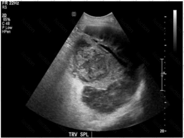
Which clinical finding is most likely associated with the splenic pathology demonstrated in this image?
A.
Trauma
B.
Sickle cell anemia
C.
Immunocompromised
D.
Portal hypertension
Full Access
Answer:
B
Explanation:
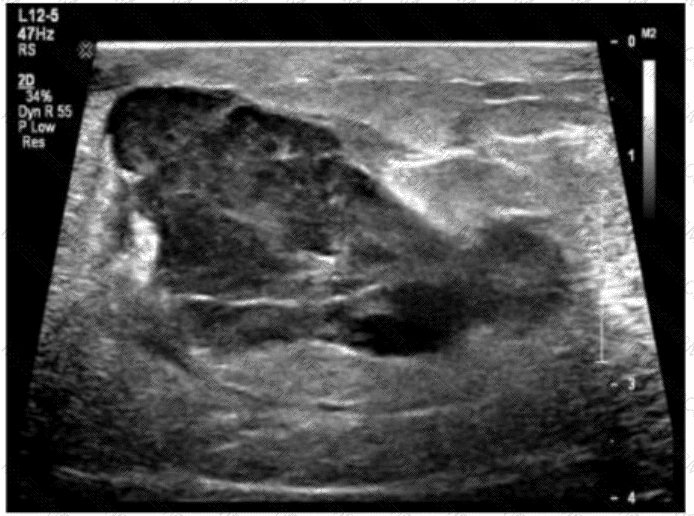
The ultrasound image demonstrates a heterogeneous and echogenic spleen with evidence of atrophy and multiple areas of calcification—consistent with autosplenectomy. This appearance is classically associated with chronic sickle cell anemia.
In sickle cell disease, repeated vaso-occlusive episodes result in infarctions, fibrosis, and progressive calcification of the spleen. Over time, this leads to functional asplenia or complete autosplenectomy (involution and shrinkage of the spleen). The hallmark sonographic features include:
A small, echogenic spleen
Multiple coarse calcifications
Irregular contour or atrophic appearance
These findings are not typically seen in other conditions:
A. Trauma may cause subcapsular hematomas or lacerations, but not chronic atrophy with calcifications.
C. Immunocompromised patients may develop abscesses or infections but not the classic features of autosplenectomy.
D. Portal hypertension typically causes splenomegaly and varices, not atrophic and calcified spleens.
[References:, Rumack CM, Wilson SR, Charboneau JW, Levine D. Diagnostic Ultrasound, 5th ed. Elsevier; 2017., Hagen-Ansert SL. Textbook of Diagnostic Sonography, 8th ed. Elsevier; 2017., Kellenberger CJ. Imaging of the spleen in children. Eur Radiol. 2004;14(5):92–102., , , ]
Question # 9
Which foreign body is better visualized with sonography than computed tomography (CT)?
A.
Glass
B.
Wood
C.
Metal
D.
Stone
Full Access
Answer:
B
Explanation:
Wooden foreign bodies are often difficult to detect on CT because of their low radiodensity, but they are highly echogenic with posterior shadowing or reverberation on ultrasound, making ultrasound superior for detecting retained wooden objects. Glass, metal, and stones are better visualized with CT due to their high radiodensity.
According to AIUM and musculoskeletal ultrasound literature:
“Wood is poorly visualized on CT but demonstrates high reflectivity and acoustic shadowing on ultrasound.â€
[Reference:, Bianchi S, Martinoli C. Ultrasound of the Musculoskeletal System. Springer, 2007., AIUM Practice Parameter for Musculoskeletal Ultrasound, 2020., —]
Question # 10
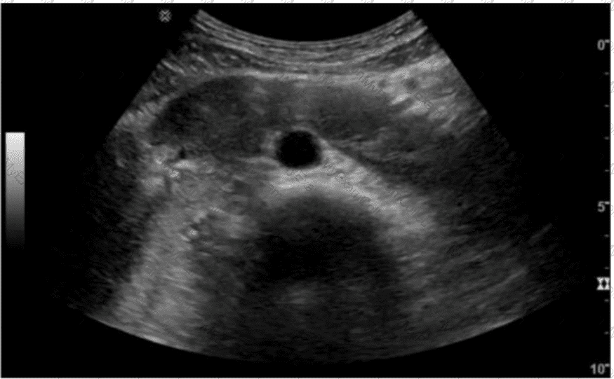
Based on this image, which congenital anomaly should be suspected?
A.
Supernumerary kidney
B.
Pancreas divisum
C.
Annular pancreas
D.
Horseshoe kidney
Full Access
Answer:
C
Explanation:
The ultrasound image demonstrates a dilated duodenum with a hypoechoic soft tissue structure encircling it. This is a classic sonographic appearance suggestive of an annular pancreas. In annular pancreas, pancreatic tissue completely or partially encircles the second portion of the duodenum, which can lead to duodenal narrowing or obstruction.
Annular pancreas is a congenital anomaly that results from failure of the ventral pancreatic bud to rotate properly during embryologic development. As a result, pancreatic tissue encircles the duodenum. It may present in neonates with symptoms of duodenal obstruction or in adults with abdominal pain, pancreatitis, or vomiting.
Ultrasound Findings:
Hypoechoic pancreatic tissue encircling the duodenum
Evidence of duodenal dilatation proximal to the obstruction
“Double bubble†sign may be seen in neonates
Differentiation from other options:
A. Supernumerary kidney: Refers to an accessory kidney. It would be seen in the retroperitoneum and is unrelated to the duodenum or pancreas.
B. Pancreas divisum: A ductal anomaly best diagnosed on MRCP or ERCP. It is not typically visible on conventional ultrasound.
D. Horseshoe kidney: A renal fusion anomaly where the lower poles of the kidneys are fused. It is seen in the pelvis or lower abdomen and does not involve the duodenum or pancreas.
[References:, Rumack CM, Wilson SR, Charboneau JW, Levine D. Diagnostic Ultrasound. 5th Edition. Elsevier, 2018. Chapter: Pancreas, pp. 269–272., Radiopaedia.org. Annular pancreas: https://radiopaedia.org/articles/annular-pancreas, AIUM Practice Parameter for the Performance of Abdominal and Retroperitoneal Ultrasound Examinations, 2020., , ]
Question # 11
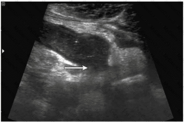
A patient presents with right lower quadrant pain and fever. Which condition is most likely indicated by the arrow on this image?
A.
Bowel obstruction
B.
Intussusception
C.
Enlarged lymph node
D.
Ruptured appendix
Full Access
Answer:
D
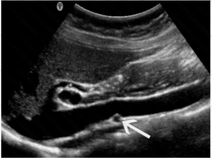
Explanation:
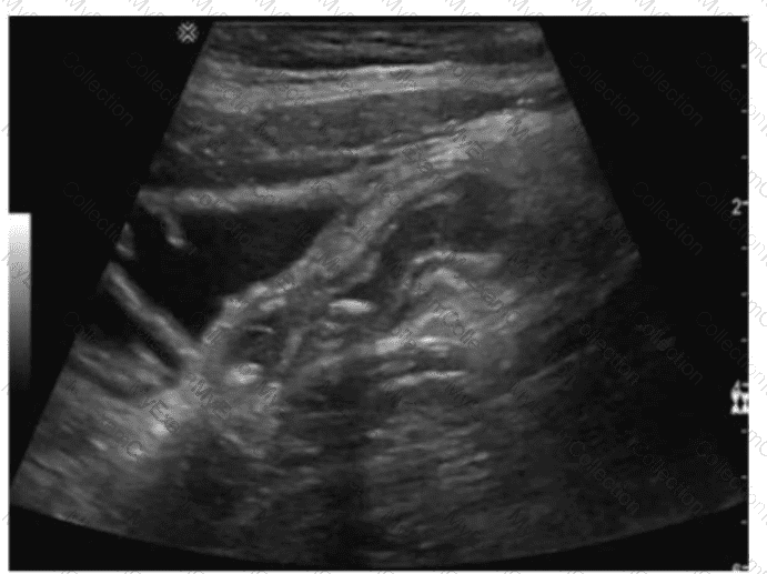
The ultrasound image demonstrates a tubular, non-compressible, blind-ending structure located in the right lower quadrant (RLQ) with associated echogenic periappendiceal fat and possibly adjacent fluid or phlegmon. These features are consistent with appendicitis. Given the clinical history of fever and RLQ pain, along with the irregular borders and complex periappendiceal findings, the diagnosis of a ruptured appendix is most likely.
Key sonographic features of ruptured appendicitis include:
Non-visualization or distortion of the normal appendiceal wall architecture
Periappendiceal fluid collection or abscess
Disruption of the echogenic submucosal layer
Surrounding fat stranding (hyperechoic inflammatory changes)
Clinical correlation with fever and peritonitis
Comparison of answer choices:
A. Bowel obstruction typically shows dilated bowel loops with air-fluid levels, not a tubular structure like the appendix.
B. Intussusception presents with a target or “donut†sign in a transverse view, not a linear tubular structure.
C. Enlarged lymph nodes are usually round or oval and hypoechoic with a central echogenic hilum, without a tubular appearance.
D. Ruptured appendix — Correct. The ultrasound features and clinical presentation match.
[References:, Rumack CM, Wilson SR, Charboneau JW, Levine D. Diagnostic Ultrasound, 5th ed. Elsevier; 2017., Jeffrey RB, Laing FC, Townsend RR. Acute appendicitis: sonographic criteria based on 250 cases. Radiology. 1988;167(2):327–329., American Institute of Ultrasound in Medicine (AIUM) Practice Parameter for the Performance of the Ultrasound Examination for Appendicitis (2020)., , ]
Question # 12
Which scanning approach was utilized to obtain this image?
A.
Anterior
B.
Posterior
C.
Left coronal
D.
Right coronal
Full Access
Answer:
D
Explanation:
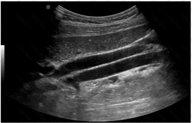
The ultrasound image provided shows the liver and diaphragm imaged in a coronal plane with characteristic rib shadows and costophrenic angles. The orientation of the image and the structures visualized suggest that the transducer is placed in the right mid-axillary line, with the sound beam directed coronally — this is a classic right coronal scanning approach.
Key features supporting this:
The liver appears superiorly in the image.
Multiple echogenic lines (representing the ribs) run obliquely, casting acoustic shadows.
The diaphragm and adjacent lung base are seen clearly, which is commonly imaged through the right intercostal spaces in a coronal plane.
Comparison of answer choices:
A. Anterior: Would show a more transverse view of the liver and not typically image the diaphragm and lung this way.
B. Posterior: Not used for upper abdominal scanning due to shadowing from the spine and posterior ribs.
C. Left coronal: Would show the spleen and left kidney — not the liver as seen here.
D. Right coronal — Correct. This image was obtained using the right coronal (intercostal) approach through the right flank.
[References:, Rumack CM, Wilson SR, Charboneau JW, Levine D. Diagnostic Ultrasound, 5th ed. Elsevier; 2017., Hagen-Ansert SL. Textbook of Diagnostic Sonography, 8th ed. Elsevier; 2017., AIUM Practice Parameter for the Performance of an Ultrasound Examination of the Abdomen and/or Retroperitoneum (2020)., , , ]
Question # 13
Which finding is expected in the contralateral kidney given the pathology depicted in this image?
A.
Duplicated collecting system
B.
Polycystic kidney
C.
Parapelvic cysts
D.
Atrophic kidney
Full Access
Answer:
B
Explanation:
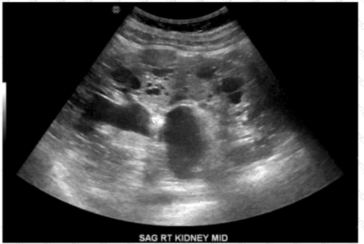
The ultrasound image shows a sagittal view of the right kidney with multiple anechoic (black), non-communicating cysts of varying sizes distributed throughout the renal parenchyma, consistent with autosomal dominant polycystic kidney disease (ADPKD).
ADPKD is a hereditary disorder characterized by the progressive development of multiple bilateral renal cysts, which leads to renal enlargement and eventual loss of function. This condition typically affects both kidneys, making bilateral polycystic involvement expected. Therefore, the same cystic appearance is anticipated in the contralateral (left) kidney as well.
Comparison of answer choices:
A. Duplicated collecting system: This is a congenital anomaly but does not result in diffusely cystic kidneys.
B. Polycystic kidney: Correct. Bilateral renal involvement is the hallmark of ADPKD.
C. Parapelvic cysts: These are simple cysts located in the renal sinus and do not exhibit the diffuse pattern seen here.
D. Atrophic kidney: Not typical in the contralateral side in ADPKD; the disease affects both kidneys symmetrically.
[References:, Rumack CM, Wilson SR, Charboneau JW, Levine D. Diagnostic Ultrasound, 5th ed. Elsevier; 2017., Hagen-Ansert SL. Textbook of Diagnostic Sonography, 8th ed. Elsevier; 2017., Torres VE, Harris PC, Pirson Y. Autosomal dominant polycystic kidney disease. Lancet. 2007;369(9569):1287–1301., , ]
Question # 14
Which best describes the Doppler waveform findings in this image?
A.
Normal
B.
Increased resistance
C.
Tardus parvus
D.
Triphasic
Full Access
Answer:
A
Explanation:
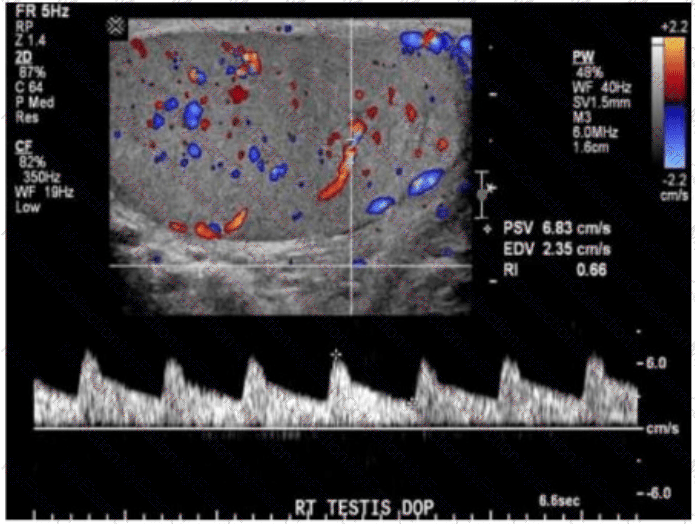
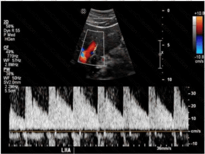
The Doppler spectral waveform shown in this image of the right testis demonstrates low-resistance, forward-flowing arterial waveforms with continuous diastolic flow — this is characteristic of normal testicular perfusion. The presence of both color Doppler flow and a resistive index (RI) of 0.66 further supports normal testicular arterial circulation.
Key Doppler features of a normal testicular waveform:
Low-resistance waveform (RI typically 0.5–0.75)
Continuous diastolic flow
No reversal of flow or spectral broadening
Color Doppler confirms uniform intratesticular vascularity
Clinical context:
Normal testicular flow on Doppler imaging excludes testicular torsion, infarction, or significant inflammation.
Testicular torsion would show either absent or very high-resistance (reduced or absent diastolic flow) waveform.
Epididymo-orchitis may show hyperemia with low resistance but often presents with other gray-scale findings like heterogeneous echotexture or scrotal wall thickening.
Differentiation from other options:
B. Increased resistance: RI >0.75 and reduced or reversed diastolic flow; may indicate impending torsion or ischemia.
C. Tardus parvus: A slow systolic upstroke and diminished amplitude; indicates proximal arterial stenosis.
D. Triphasic: Normal waveform in peripheral arteries, such as extremities, not seen in testicular circulation.
[References:, Rumack CM, Wilson SR, Charboneau JW, Levine D. Diagnostic Ultrasound. 5th Edition. Elsevier, 2018. Chapter: Male Pelvis – Testis and Scrotum, pp. 793–800., AIUM Practice Parameter for the Performance of Scrotal Ultrasound Examinations, 2020., Radiopaedia.org. Testicular Doppler assessment: https://radiopaedia.org/articles/testicular-doppler-assessment, , ]
Question # 15
What is the location of the left lobe of the thyroid gland?
A.
Anterior to the left jugular vein
B.
Posterior to the longus colli muscle
C.
Anterolateral to the esophagus
D.
Anterior to the trachea
Full Access
Answer:
C
Explanation:
The left lobe of the thyroid is located anterolateral to the esophagus. On transverse ultrasound imaging, the esophagus can often be seen posterior to the left thyroid lobe as a circular structure with echogenic mucosa and hypoechoic muscular layer. The longus colli muscle lies posterior to the thyroid. The thyroid is anterior to the trachea but this refers more to the isthmus or midline portion.
According to Rumack’s Diagnostic Ultrasound:
“The esophagus is seen as a target-shaped structure posterior to the left thyroid lobe; thus, the thyroid lobe is anterolateral to the esophagus.â€
[Reference:, Rumack CM, Wilson SR, Charboneau JW, Levine D. Diagnostic Ultrasound. 5th ed. Elsevier, 2017., AIUM Practice Parameter for Thyroid Ultrasound, 2020., —]
Question # 16
Which condition is most consistent with thinning of the renal cortex, reduction in renal length, and prominence of the renal sinus fat in a patient presenting four months after renal transplant with slightly reduced renal function?
A.
Acute rejection
B.
Chronic rejection
C.
Normal findings
D.
Arterial stricture
Full Access
Answer:
B
Explanation:
Chronic rejection presents sonographically as cortical thinning, decreased renal size, and increased echogenicity of the renal sinus fat. Acute rejection typically causes an enlarged, edematous kidney with increased parenchymal echogenicity but preserved size early on.
According to Zwiebel’s Introduction to Vascular Ultrasound:
“In chronic rejection, the allograft becomes smaller with cortical thinning, increased echogenicity, and prominence of the central sinus fat.â€
[Reference:, Zwiebel WJ, Pellerito JS. Introduction to Vascular Ultrasound. 6th ed. Elsevier, 2019., AIUM Practice Parameter for Renal Transplant Ultrasound, 2020., —]
Question # 17
Which structure is indicated by the arrow on this image?
A.
Parathyroid
B.
Lymph node
C.
Esophagus
D.
Paraganglioma
Full Access
Answer:
C
Explanation:
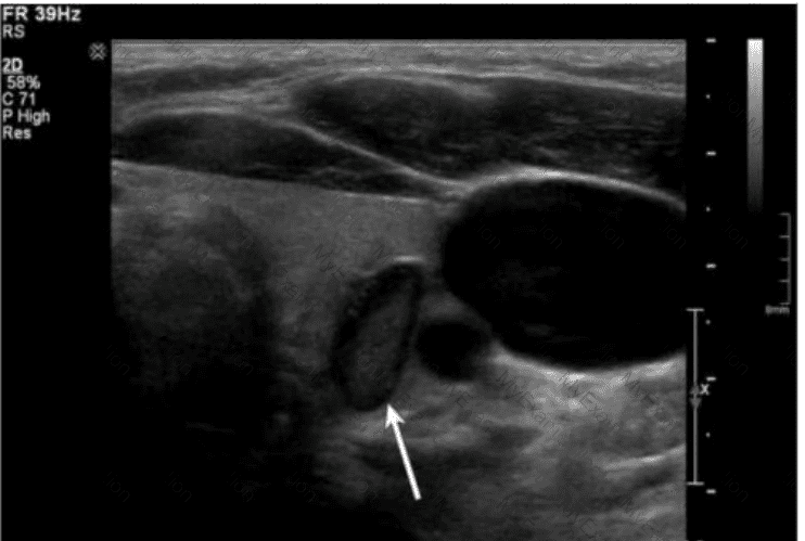
The ultrasound image shows a transverse view of the lower neck region at the thyroid level. The arrow is pointing to a round-to-oval structure located posterior and slightly to the left of the thyroid gland. The structure has a characteristic "target" or "bull's-eye" appearance with a hypoechoic outer ring and echogenic central mucosal interface — this is classic for the esophagus when seen in transverse view.
Key sonographic features of the esophagus:
It lies posterior to the left lobe of the thyroid.
It demonstrates a layered wall structure ("target" or "bull's-eye" appearance).
It may change shape or move during swallowing, and occasionally air bubbles or movement of fluid may be observed.
Comparison of answer choices:
A. Parathyroid glands are small, homogeneous, hypoechoic, and located posterior to the thyroid — but do not have this layered target appearance.
B. Lymph nodes have a hypoechoic cortex and echogenic hilum and are typically oval or bean-shaped, without the concentric ring appearance.
C. Esophagus — Correct. The location, appearance, and structure are consistent with the cervical esophagus.
D. Paragangliomas are highly vascular and more commonly located in the carotid body or adrenal region, not in this location or with this sonographic pattern.
[References:, Rumack CM, Wilson SR, Charboneau JW, Levine D. Diagnostic Ultrasound, 5th ed. Elsevier; 2017., Grant EG, Tessler FN, Hoang JK, et al. Thyroid Ultrasound Reporting Lexicon: White Paper of the ACR TI-RADS Committee. J Am Coll Radiol. 2015., Hagen-Ansert SL. Textbook of Diagnostic Sonography, 8th ed. Elsevier; 2017., , , ]
Question # 18
What is the most common cause of nutcracker syndrome?
A.
Compression of left renal vein between inferior vena cava and aorta
B.
Compression of left renal vein between superior mesenteric artery and aorta
C.
Compression of right renal vein between superior mesenteric artery and aorta
D.
Compression of right renal vein between inferior vena cava and aorta
Full Access
Answer:
B
Explanation:
Nutcracker syndrome results from compression of the left renal vein between the superior mesenteric artery (SMA) and the aorta. This can cause hematuria, flank pain, and pelvic congestion due to impaired venous drainage.
According to Zwiebel’s Introduction to Vascular Ultrasound:
“In nutcracker syndrome, the left renal vein is compressed between the aorta and SMA, resulting in venous hypertension.â€
[Reference:, Zwiebel WJ, Pellerito JS. Introduction to Vascular Ultrasound. 6th ed. Elsevier, 2019., AIUM Practice Parameter for Abdominal Vascular Ultrasound, 2020., —]
Question # 19
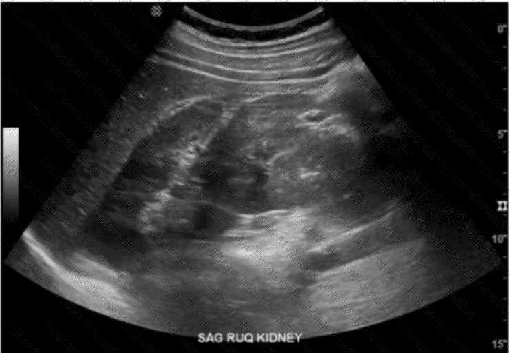
Which renal anomaly is demonstrated on this image?
A.
Duplicated collecting system
B.
Crossed renal ectopia
C.
Horseshoe kidney
D.
Pelvic kidney
Full Access
Answer:
C
Explanation:
The ultrasound image labeled “SAG RUQ KIDNEY†demonstrates a midline sagittal view showing a renal parenchymal structure that extends across the midline anterior to the aorta and vertebral bodies, suggesting the presence of a horseshoe kidney.
A horseshoe kidney is a congenital renal anomaly in which the lower poles of both kidneys are fused across the midline by a parenchymal or fibrous isthmus. This isthmus typically lies anterior to the aorta and inferior vena cava and can be seen as a hypoechoic band of tissue crossing the midline on ultrasound.
Ultrasound findings characteristic of a horseshoe kidney:
Abnormally low position of the kidneys in the abdomen
Renal tissue (isthmus) bridging the lower poles anterior to the great vessels
Renal axes may be more horizontal than usual
Kidneys may appear closer together or “kissing†the spine anteriorly
Differentiation from other options:
A. Duplicated collecting system: Manifests as two separate collecting systems within one kidney, often with a central renal sinus split into two — not typically midline bridging.
B. Crossed renal ectopia: Involves one kidney crossing midline and fusing with the other on the opposite side, but they do not form a midline isthmus.
D. Pelvic kidney: A single kidney located in the pelvis due to failed ascent — it does not appear as midline fusion of two kidneys.
[References:, Rumack CM, Wilson SR, Charboneau JW, Levine D. Diagnostic Ultrasound. 5th Edition. Elsevier, 2018. Chapter: Urinary Tract, pp. 215–218., American Institute of Ultrasound in Medicine (AIUM). Practice Parameter for the Performance of an Ultrasound Examination of the Abdomen and/or Retroperitoneum. 2020., Radiopaedia.org. Horseshoe kidney: https://radiopaedia.org/articles/horseshoe-kidney, , , ]
Question # 20
Which complication is of greatest concern with undescended testis?
A.
Hydrocele
B.
Seminoma
C.
Torsion
D.
Hernia
Full Access
Answer:
B
Explanation:
The most serious long-term complication of undescended testis (cryptorchidism) is an increased risk of testicular malignancy, especially seminoma. Although torsion and hernia may also occur, seminoma is the most concerning complication due to its life-threatening potential.
According to Rumack’s Diagnostic Ultrasound:
“Cryptorchidism is associated with a significantly increased risk of seminoma, the most common malignancy in undescended testes.â€
[Reference:, Rumack CM, Wilson SR, Charboneau JW, Levine D. Diagnostic Ultrasound. 5th ed. Elsevier, 2017., American Urological Association (AUA) Guidelines, 2019., —]
Question # 21
Which action should a sonographer take if the abdominal aorta measures 5.5 centimeters in the anteroposterior diameter?
A.
Report the finding to the radiologist immediately
B.
Follow the routine protocol for abdominal ultrasound
C.
Release patient from care
D.
Disclose the diagnosis to the patient
Full Access
Answer:
A
Explanation:
An abdominal aortic aneurysm (AAA) measuring ≥5.5 cm represents a significantly increased risk of rupture and often requires surgical evaluation. The sonographer must report this critical finding immediately to the interpreting physician. The sonographer should never disclose a diagnosis directly to the patient.
According to AIUM and SRU Guidelines:
“An aortic diameter of 5.5 cm or greater should be promptly reported to the interpreting physician due to the high risk of rupture.â€
[Reference:, AIUM Practice Parameter for Abdominal Aortic Ultrasound, 2020., Society of Radiologists in Ultrasound (SRU) Consensus Statement, 2003.]
Question # 22
Which thyroid condition is most likely caused by a viral infection?
A.
Hashimoto
B.
Graves
C.
Abscess
D.
De Quervain
Full Access
Answer:
D
Explanation:
De Quervain thyroiditis (subacute granulomatous thyroiditis) is often triggered by a viral infection. Patients may present with painful thyroid enlargement, elevated inflammatory markers, and transient hyperthyroidism. Hashimoto’s and Graves' diseases are autoimmune in nature.
According to Braverman’s The Thyroid:
“Subacute (De Quervain) thyroiditis typically follows a viral upper respiratory tract infection and is characterized by thyroid pain and transient thyrotoxicosis.â€
[Reference:, Braverman LE, Cooper DS. The Thyroid: A Fundamental and Clinical Text. 11th ed. Wolters Kluwer, 2021., American Thyroid Association Guidelines, 2016.]
Question # 23
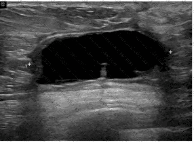
Which condition is most consistent with this image of a postsurgical breast?
A.
Carcinoma
B.
Blood clot
C.
Abscess
D.
Seroma
Full Access
Answer:
D
Explanation:
The ultrasound image reveals a well-defined, anechoic (black), thin-walled fluid collection located in the subcutaneous or parenchymal plane of the breast. This is most consistent with a seroma, particularly in the context of recent breast surgery.
A seroma is a common postsurgical finding, representing a sterile collection of serous fluid that accumulates in the surgical bed. It typically appears:
Anechoic (or hypoechoic if older)
Well circumscribed
Without internal septations or debris
Lacking hyperemia or surrounding inflammatory changes
This contrasts with:
A. Carcinoma — typically presents as an irregular, hypoechoic mass with angular margins, internal vascularity, and shadowing.
B. Blood clot (hematoma) — often appears heterogeneous, with internal echoes and variable echotexture depending on the age of the clot.
C. Abscess — appears as a complex fluid collection with thick walls, internal debris, septations, and surrounding hyperemia (often with clinical signs of infection).
D. Seroma — Correct. The described anechoic, clean-walled fluid collection is classic for a postoperative seroma.
[References:, Mendelson EB, Böhm-Vélez M, Berg WA.ACR BI-RADS® Atlas: Ultrasound. American College of Radiology; 2013., Stavros AT. Breast Ultrasound. Lippincott Williams & Wilkins; 2004., Rumack CM, Wilson SR, Charboneau JW, Levine D. Diagnostic Ultrasound, 5th ed. Elsevier; 2017., , ]
Question # 24
Which syndrome is characterized by right upper quadrant pain, ascites, and hepatocellular dysfunction?
A.
Budd-Chiari
B.
Calciphylaxis
C.
Ehlers-Danlos
D.
Klippel-Trenaunay
Full Access
Answer:
A
Explanation:
Budd-Chiari syndrome is caused by hepatic venous outflow obstruction, resulting in hepatomegaly, ascites, right upper quadrant pain, and liver dysfunction. It may be due to thrombosis or compression of the hepatic veins or IVC.
According to Rumack’s Diagnostic Ultrasound:
“Budd-Chiari syndrome results from hepatic venous outflow obstruction and presents with hepatomegaly, ascites, and right upper quadrant pain.â€
[Reference:, Rumack CM, Wilson SR, Charboneau JW, Levine D. Diagnostic Ultrasound. 5th ed. Elsevier, 2017., AIUM Practice Parameter for Liver Ultrasound, 2020., —]
Question # 25
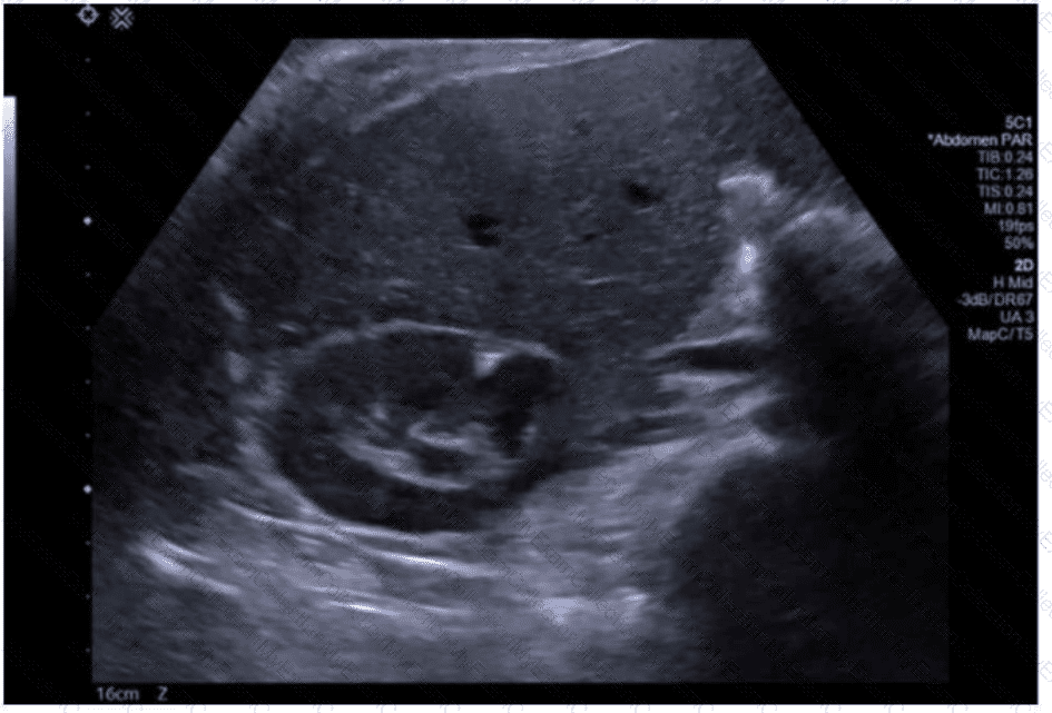
Identify the congenital anomaly.
Using your mouse, place the cursor on the appropriate region of the image and then left-click the mouse button to indicate your selection.
Full Access
Answer:
Answer: 
Explanation:
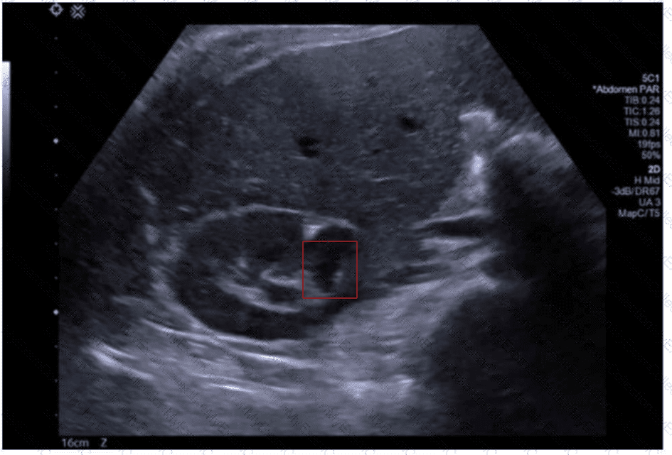 An ultrasound of a fetus
AI-generated content may be incorrect.
An ultrasound of a fetus
AI-generated content may be incorrect.
The ultrasound image shows a transverse (axial) view of the fetal abdomen. Notably, there is abnormal continuity of renal parenchyma across the midline anterior to the aorta, forming a U- or horseshoe-shaped structure. This is characteristic of a congenital anomaly known as a horseshoe kidney.
Horseshoe kidney is the most common fusion anomaly of the kidneys, occurring in approximately 1 in 400–600 live births. It results from fusion of the lower poles of both kidneys during fetal development. On prenatal ultrasound, this anomaly can be suspected when the kidneys appear closer to the midline than usual and are connected by an isthmus of renal tissue or fibrous band that crosses anterior to the spine and great vessels.
Typical sonographic findings include:
Abnormally located kidneys, often lower than expected
Renal fusion across the midline (usually at the lower poles)
Possible associated hydronephrosis or malrotation
Comparison to other anomalies:
This is not consistent with polycystic kidney disease (which would show diffusely echogenic kidneys with poor corticomedullary differentiation).
Duplex kidney would show duplicated collecting systems but not fusion across the midline.
Renal agenesis would demonstrate absence of renal tissue.
Posterior urethral valves would show a distended bladder with bilateral hydronephrosis, not midline fusion.
[References:, Rumack CM, Wilson SR, Charboneau JW, Levine D. Diagnostic Ultrasound, 5th ed. Elsevier; 2017., Callen PW. Ultrasonography in Obstetrics and Gynecology, 6th ed. Elsevier; 2016., Nyberg DA, McGahan JP, Pretorius DH, Pilu G. Diagnostic Imaging of Fetal Anomalies. Lippincott Williams & Wilkins; 2003., , , ]
Question # 26
Which condition results in the vascular abnormality shown in this image of a renal transplant?
A.
Iliac arteritis
B.
Renal artery stenosis
C.
Renal vein thrombosis
D.
Arteriovenous malformation
Full Access
Answer:
B
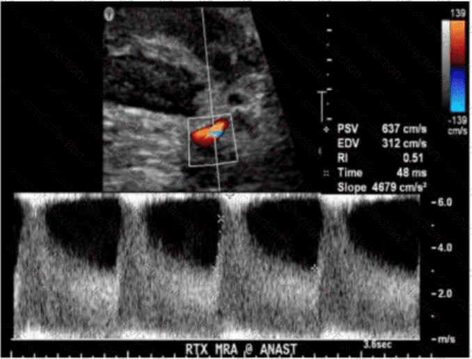
Explanation:
The Doppler ultrasound image shows an elevated peak systolic velocity (PSV) of 637 cm/s, an elevated end-diastolic velocity (EDV) of 312 cm/s, and a low resistive index (RI) of 0.51 at the arterial anastomosis of a renal transplant. These findings are characteristic of significant renal artery stenosis (RAS) at the transplant vascular anastomosis.
Key sonographic features of renal artery stenosis:
Peak systolic velocity (PSV) > 250–300 cm/s at the stenotic segment (this case: 637 cm/s)
Post-stenotic turbulence with spectral broadening
Low resistive index (RI < 0.56 suggests downstream vasodilation)
Elevated acceleration time (AT > 0.07 sec), and reduced acceleration slope
Aliasing on color Doppler due to high velocity
In this image, the marked increase in velocity with spectral aliasing and low RI is diagnostic of transplant renal artery stenosis — the most common vascular complication post-transplant, typically occurring at the site of surgical anastomosis.
Differentiation from other options:
A. Iliac arteritis: A rare condition, not typically presenting with these Doppler changes.
C. Renal vein thrombosis: Would show reversed or absent diastolic flow, not elevated systolic velocities.
D. Arteriovenous malformation (AVM): Produces a high-velocity, low-resistance waveform but is associated with color bruit, aliasing, and pulsatile venous waveforms — not evident here.
[References:, Rumack CM, Wilson SR, Charboneau JW, Levine D. Diagnostic Ultrasound. 5th Edition. Elsevier, 2018. Chapter: Transplant Imaging, pp. 1035–1045., American Institute of Ultrasound in Medicine (AIUM). Practice Parameter for the Performance of a Renal Artery Duplex Sonographic Examination, 2020., Radiopaedia.org. Renal artery stenosis (transplant): https://radiopaedia.org/articles/renal-artery-stenosis-transplant, , , ]
Question # 27
Which vessel is located directly proximal to the origination of the renal arteries?
A.
Left portal vein
B.
Splenic vein
C.
Hepatic artery
D.
Superior mesenteric artery
Full Access
Answer:
D
Explanation:
The renal arteries originate from the abdominal aorta just inferior to the superior mesenteric artery (SMA). The SMA arises anteriorly from the abdominal aorta at the level of L1, and just below it, the renal arteries branch laterally. The splenic vein, portal vein, and hepatic artery are located more superiorly in relation to the renal arteries.
According to Moore's Clinically Oriented Anatomy:
"The superior mesenteric artery arises from the anterior surface of the abdominal aorta just above the renal arteries." (Moore KL et al., Clinically Oriented Anatomy, 8th ed.)
[Reference:, Moore KL, Dalley AF, Agur AMR. Clinically Oriented Anatomy. 8th ed. Wolters Kluwer, 2018., Gray’s Anatomy for Students, 4th ed., Elsevier, 2019., ]
Question # 28
Which term best describes the common bile duct measured in this image of a postcholecystectomy patient?

A.
Normal
B.
Dilated
C.
Inflamed
D.
Atretic
Full Access
Answer:
A
Explanation:
The ultrasound image shows a measured common bile duct (CBD) diameter of 7.9 mm in a postcholecystectomy patient. In patients who have undergone cholecystectomy, mild dilation of the CBD is considered normal and is a well-recognized post-surgical change.
Normal upper limits for CBD diameter:
In patients with a gallbladder: ≤6 mm is generally considered normal.
In postcholecystectomy patients: up to 10 mm is considered within normal limits, as the CBD compensates for the absence of the gallbladder and slightly enlarges over time.
With aging, the CBD may enlarge by approximately 1 mm per decade after age 60.
Therefore, a CBD diameter of 7.9 mm in a patient without a gallbladder is considered normal.
Differentiation from other options:
B. Dilated: This would typically refer to a CBD diameter >10 mm in postcholecystectomy patients, or >6 mm in patients with an intact gallbladder.
C. Inflamed: Inflammation refers to wall thickening or hyperemia, which is not evaluated simply by measuring diameter.
D. Atretic: Describes a congenitally absent or severely narrowed duct — not applicable here.
[References:, Rumack CM, Wilson SR, Charboneau JW, Levine D. Diagnostic Ultrasound. 5th Edition. Elsevier, 2018. Chapter: Biliary System, pp. 143–146., American Institute of Ultrasound in Medicine (AIUM) Practice Parameter for the Performance of a Hepatobiliary Ultrasound Examination, 2020., Radiopaedia.org. Common bile duct: https://radiopaedia.org/articles/common-bile-duct, ]
Question # 29
Where is the most common location for a branchial cyst in relation to the thyroid?
A.
Lateral
B.
Medial
C.
Anterior
D.
Posterior
Full Access
Answer:
A
Explanation:
Branchial cleft cysts are congenital epithelial cysts that typically occur laterally in the neck, often anterior to the sternocleidomastoid muscle, and lateral to the thyroid gland. The second branchial cleft cyst is the most common type and is found in the lateral neck region.
Medial (B) would be more consistent with thyroglossal duct cysts.
Anterior (C) or posterior (D) do not specifically describe branchial cyst location relative to the thyroid.
Reference Extracts:
Som PM, Curtin HD. Head and Neck Imaging. 5th ed. Elsevier, 2011.
Rumack CM, Wilson SR, Charboneau JW, Levine D. Diagnostic Ultrasound. 5th ed. Elsevier, 2017.
—
Question # 30
Which technique best differentiates a bladder mass from a hematoma?
A.
Use harmonic imaging
B.
Obtain post-void image
C.
Fill the bladder
D.
Change patient position
Full Access
Answer:
D
Explanation:
Changing the patient's position allows evaluation of lesion mobility. Blood clots and hematomas are often mobile, while true bladder wall masses remain fixed. This technique helps differentiate between solid masses and non-adherent debris.
According to Rumack’s Diagnostic Ultrasound:
“Changing patient position may distinguish between mobile blood clots and fixed bladder wall masses.â€
[Reference:, Rumack CM, Wilson SR, Charboneau JW, Levine D. Diagnostic Ultrasound. 5th ed. Elsevier, 2017., AIUM Practice Parameter for Bladder Ultrasound, 2020., ]
Question # 31
Which retroperitoneal finding is most likely associated with trauma?
A.
Neuroblastoma
B.
Fibrosis
C.
Urinoma
D.
Adenoma
Full Access
Answer:
C
Explanation:
Urinomas are collections of urine in the retroperitoneum that result from trauma, surgery, or obstruction causing urine leakage. Trauma is a frequent cause of urinoma formation due to disruption of the urinary tract.
According to Rumack’s Diagnostic Ultrasound:
“Urinomas may develop as a complication of trauma, surgery, or obstructive uropathy with urinary extravasation into the retroperitoneum.â€
[Reference:, Rumack CM, Wilson SR, Charboneau JW, Levine D. Diagnostic Ultrasound. 5th ed. Elsevier, 2017., AIUM Practice Parameter for Renal Ultrasound, 2020., —]
Question # 32
Which finding is most likely demonstrated in this abdominal wall image of a patient with a history of atrial fibrillation?
A.
Hernia
B.
Lipoma
C.
Abscess
D.
Hematoma
Full Access
Answer:
D
Explanation:
The ultrasound image demonstrates a complex, heterogeneous hypoechoic collection within the abdominal wall, with mixed echogenicity and ill-defined margins. The lesion appears to contain internal debris but lacks definitive signs of vascularity or air (which would be seen in an abscess). There is no peristalsis, herniated bowel, or fat to suggest hernia.
Given the history of atrial fibrillation — a condition commonly treated with anticoagulation therapy (e.g., warfarin, apixaban) — this clinical background raises high suspicion for a rectus sheath or abdominal wall hematoma.
Key ultrasound features of hematomas:
Early (acute): hyperechoic or heterogeneous
Chronic/resolving: complex or cystic with fluid-debris levels
No internal vascularity on Doppler
May be confined to muscle or fascial planes
This is consistent with a hematoma, particularly in patients on anticoagulation therapy.
Comparison of answer choices:
A. Hernia — typically shows bowel or fat with movement/peristalsis passing through a fascial defect.
B. Lipoma — usually homogeneous and echogenic, not complex or fluid-filled.
C. Abscess — often presents as a complex fluid collection with peripheral hyperemia and possibly air, plus systemic signs of infection.
D. Hematoma — Correct. The image and clinical history (anticoagulation due to atrial fibrillation) strongly support this diagnosis.
[References:, Berman L, et al. Sonographic appearance and evolution of rectus sheath hematomas. AJR Am J Roentgenol. 1996., Rumack CM, Wilson SR, Charboneau JW, Levine D. Diagnostic Ultrasound, 5th ed. Elsevier; 2017., AIUM Practice Parameter for the Performance of Diagnostic Ultrasound Examinations of the Abdomen and Retroperitoneum (2020)., ]
Question # 33
Which probe frequency is most appropriate for imaging of the salivary glands?
A.
2 MHz
B.
4 MHz
C.
8 MHz
D.
12 MHz
Full Access
Answer:
D
Explanation:
Salivary glands are superficial structures, and high-frequency transducers (10–15 MHz) are optimal to obtain high spatial resolution. Lower frequencies are inappropriate as they lack sufficient resolution for superficial structures. A 12 MHz transducer provides excellent detail necessary for detecting small lesions, duct abnormalities, and vascular structures.
According to Rumack et al., Diagnostic Ultrasound:
"High-frequency linear transducers (10–15 MHz) are recommended for evaluating superficial structures such as salivary glands." (Rumack CM et al., Diagnostic Ultrasound, 5th ed.).
[Reference:, Rumack CM, Wilson SR, Charboneau JW, Levine D. Diagnostic Ultrasound. 5th ed. Elsevier; 2017., AIUM Practice Parameter for the Performance of a Head and Neck Ultrasound Examination, 2020., —]
Question # 34
Which structure is most likely shown in this image of the right lower quadrant?
A.
Fallopian tube
B.
Ureter
C.
Appendix
D.
Jejunum
Full Access
Answer:
C
Explanation:
The ultrasound image shows a blind-ending, non-compressible, tubular structure in the right lower quadrant with a target or bullseye appearance in transverse section — highly suggestive of the appendix.
Sonographic features of the appendix (especially in suspected appendicitis):
Blind-ending tubular structure arising from the cecum
Non-compressible on graded compression
Diameter >6 mm is suggestive of appendicitis
May demonstrate a “target sign†in transverse view (concentric ring-like appearance)
Increased echogenicity of surrounding fat in cases of inflammation
May contain an appendicolith or show hyperemia on color Doppler if inflamed
The location (right lower quadrant) and appearance in this case are classic for the normal or potentially inflamed appendix.
Differentiation from other options:
A. Fallopian tube: Located more in the adnexal regions and usually not visible unless distended (e.g., hydrosalpinx).
B. Ureter: Usually not visualized on ultrasound unless dilated due to obstruction.
D. Jejunum: Has valvulae conniventes ("keyboard sign") and peristalsis; does not present with a blind-ending tubular appearance from the cecum.
[References:, Rumack CM, Wilson SR, Charboneau JW, Levine D. Diagnostic Ultrasound. 5th Edition. Elsevier, 2018. Chapter: Gastrointestinal Tract, pp. 460–468., American College of Radiology (ACR). ACR Appropriateness Criteria® — Right Lower Quadrant Pain – Suspected Appendicitis., AIUM Practice Parameter for the Performance of a Pediatric Abdominal and/or Retroperitoneal Ultrasound Examination, 2020., , ]
Question # 35
Which outcome would be present if the sample volume gate is larger than the examined vessel?
A.
Indeterminate flow direction
B.
Spike turbulence
C.
Spectral noise
D.
Aliasing
Full Access
Answer:
C
Explanation:
When the sample volume (gate) is too large, it captures signals from both the vessel and surrounding tissues or adjacent flows. This leads to a broadening of the spectral waveform and produces "spectral noise" or "spectral broadening," reducing the accuracy of velocity measurements and waveform analysis. Aliasing results from high velocity relative to the Nyquist limit, not from gate size.
According to Zwiebel’s Introduction to Vascular Ultrasound:
“Increasing the sample volume beyond the vessel size causes spectral broadening, resulting in spectral noise and inaccurate Doppler measurements.â€
[Reference:, Zwiebel WJ, Pellerito JS. Introduction to Vascular Ultrasound. 6th ed. Elsevier, 2019., AIUM Practice Parameter for Spectral Doppler Ultrasound, 2021., —]
Question # 36
During a renal artery Doppler study, which vessel should also be sampled to verify patency?
A.
Inferior vena cava
B.
Main renal vein
C.
Portal vein
D.
Iliac vein
Full Access
Answer:
B
Explanation:
The main renal vein should be assessed in addition to the renal arteries during renal Doppler exams. Venous thrombosis may coexist with arterial abnormalities and can impact renal perfusion. Evaluation of both arterial inflow and venous outflow ensures a comprehensive assessment of renal vascular patency.
According to Zwiebel’s Introduction to Vascular Ultrasound:
“Renal vein assessment should be performed during renal artery Doppler studies to exclude venous thrombosis or outflow obstruction.â€
[Reference:, Zwiebel WJ, Pellerito JS. Introduction to Vascular Ultrasound. 6th ed. Elsevier, 2019., AIUM Practice Parameter for Renal Artery Duplex Sonography, 2020., —]
Question # 37
Which normal anatomical structure is also known as the accessory pancreatic duct?
A.
Duct of Vater
B.
Duct of Wirsung
C.
Duct of Santorini
D.
Common pancreatic duct
Full Access
Answer:
C
Explanation:
The Duct of Santorini is the accessory pancreatic duct that drains the superior portion of the pancreatic head into the minor duodenal papilla. The main pancreatic duct (Duct of Wirsung) drains into the major papilla, often joining the common bile duct at the Ampulla of Vater.
According to Moore’s Clinically Oriented Anatomy:
“The accessory pancreatic duct (Duct of Santorini) may be present and drains into the minor duodenal papilla.â€
[Reference:, Moore KL, Dalley AF, Agur AMR. Clinically Oriented Anatomy. 8th ed. Wolters Kluwer, 2018., Gray’s Anatomy for Students, 4th ed., Elsevier, 2019., —]
Question # 38
Which characteristic is associated with complex pleural effusion?
A.
Homogeneous hypoechoic
B.
Anechoic without locules
C.
Dependent layering echoes
D.
Contains septa
Full Access
Answer:
D
Explanation:
A complex pleural effusion often contains internal septations or fibrin strands, distinguishing it from simple anechoic effusion. These septations suggest exudative processes such as infection, malignancy, or hemothorax.
According to Rumack’s Diagnostic Ultrasound:
“Complex pleural effusions demonstrate internal septations or loculations, often related to infection or malignancy.â€
[Reference:, Rumack CM, Wilson SR, Charboneau JW, Levine D. Diagnostic Ultrasound. 5th ed. Elsevier, 2017., AIUM Practice Parameter for Thoracic Ultrasound, 2020., —]
Question # 39
Which adjustment would most likely improve visualization of a small superficial tubular structure such as a peripheral artery?
A.
Decreasing frame rate
B.
Decreasing transducer wavelength
C.
Decreasing slice width
D.
Decreasing power output
Full Access
Answer:
C
Explanation:
Reducing slice (section) width improves spatial resolution, particularly elevational resolution, which enhances visualization of small, superficial structures. Lower slice width reduces off-axis beam artifacts and blurring. Wavelength depends on transducer frequency, not adjustable directly during scanning.
According to Zwiebel’s Introduction to Vascular Ultrasound:
“Reduction in slice thickness improves imaging of small superficial structures by minimizing volume averaging and improving elevational resolution.â€
[Reference:, Zwiebel WJ, Pellerito JS. Introduction to Vascular Ultrasound. 6th ed. Elsevier, 2019., AIUM Practice Parameter for Vascular Ultrasound, 2021., —]
Question # 40
Which complication would be associated with retroperitoneal fibrosis?
A.
Aortic stenosis
B.
Portal hypertension
C.
Venous thrombosis
D.
Hydronephrosis
Full Access
Answer:
D
Explanation:
Retroperitoneal fibrosis can encase and compress the ureters, leading to obstructive uropathy and hydronephrosis. It may also involve other retroperitoneal structures but hydronephrosis is the most common significant complication.
According to Rumack’s Diagnostic Ultrasound:
“Retroperitoneal fibrosis frequently results in ureteral obstruction, leading to hydronephrosis.â€
[Reference:, Rumack CM, Wilson SR, Charboneau JW, Levine D. Diagnostic Ultrasound. 5th ed. Elsevier, 2017., AIUM Practice Parameter for Abdominal Ultrasound, 2020., —]
Question # 41
Which description best characterizes a normal systolic spectral waveform of the renal artery?
A.
Slow acceleration
B.
Blunt peak
C.
Early reversal
D.
Rapid acceleration
Full Access
Answer:
D
Explanation:
A normal renal artery waveform demonstrates rapid systolic upstroke (acceleration) with continuous forward flow in diastole due to the kidney's low-resistance vascular bed. Slow acceleration or blunted peaks may indicate significant renal artery stenosis.
According to Zwiebel’s Introduction to Vascular Ultrasound:
“Normal renal artery waveforms demonstrate a rapid systolic acceleration with a sharp systolic peak.â€
[Reference:, Zwiebel WJ, Pellerito JS. Introduction to Vascular Ultrasound. 6th ed. Elsevier, 2019., ACR Practice Parameter for the Performance of a Duplex Doppler Examination, 2021., —]
Question # 42
Which hernia characteristic is demonstrated in these images?
A.
Fat only
B.
Reducible
C.
Incarcerated
D.
Strangulated
Full Access
Answer:
B
Explanation:
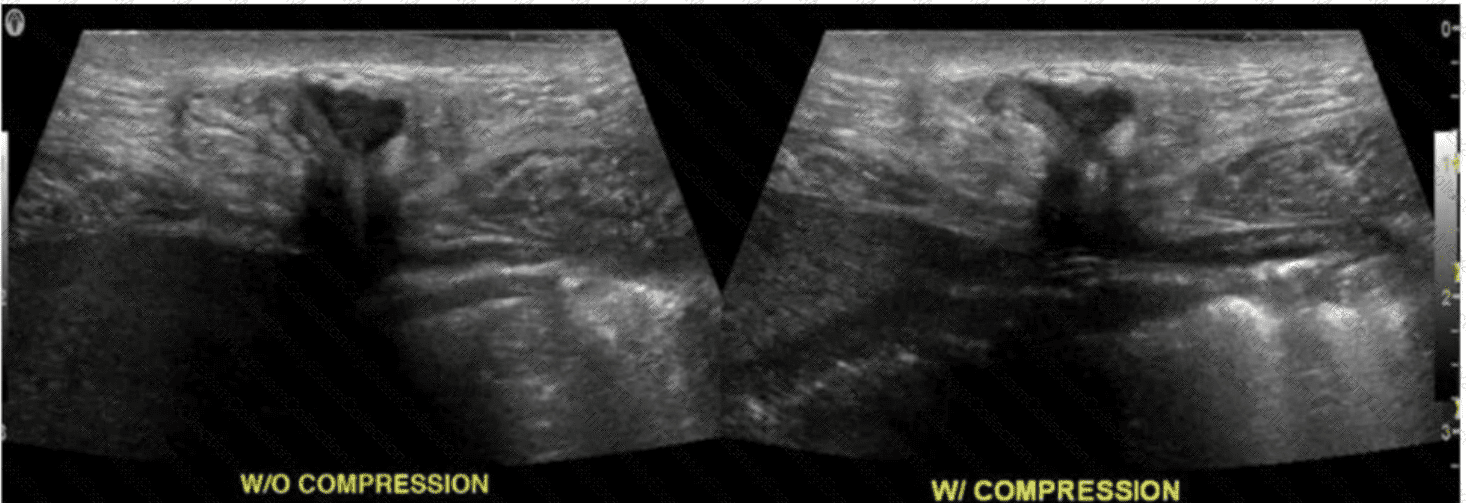
The ultrasound images show two views of the same groin region — one without compression (left image labeled “W/O COMPRESSIONâ€) and one with graded probe compression (right image labeled “W/ COMPRESSIONâ€).
In the non-compression image, a hypoechoic mass-like structure is visible protruding through the abdominal wall, consistent with a hernia sac. On the compression image, the herniated content is no longer visible, indicating that the contents have been pushed back into the abdominal cavity. This is the hallmark feature of a reducible hernia.
Key characteristics of a reducible hernia on ultrasound:
Herniated contents are visible without pressure.
Contents disappear or reduce back into the abdomen with graded probe compression or Valsalva release.
Typically includes omental fat or bowel, but reduction confirms lack of incarceration or strangulation.
Comparison of answer choices:
A. Fat only refers to the hernia content type, not the behavior or reducibility shown here.
B. Reducible — Correct. The change in hernia appearance between images demonstrates successful reduction with compression.
C. Incarcerated hernia would remain visible and not compressible or reducible.
D. Strangulated hernia would show signs of ischemia (bowel wall thickening, absent perfusion, hyperechoic mesentery), and would also not reduce with compression.
[References:, Radswiki. Ultrasound evaluation of hernia. Radiopaedia.org, Rumack CM, Wilson SR, Charboneau JW, Levine D. Diagnostic Ultrasound, 5th ed. Elsevier; 2017., AIUM Practice Parameter for the Performance of a Focused Ultrasound Examination for Hernia (2021), , , ]
Question # 43
Which congenital disorder is most consistent with the finding identified by the arrow on this image?
A.
Sclerosing cholangitis
B.
Alagille syndrome
C.
Caroli disease
D.
Biliary atresia
Full Access
Answer:
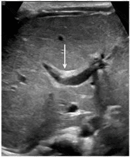
C
Explanation:
The image demonstrates a characteristic "central dot sign" — a hallmark finding of Caroli disease. This is best appreciated on ultrasound as a cystic dilation of the intrahepatic bile ducts with a central echogenic dot or linear structure (which corresponds to the portal vein and fibrous tissue within the dilated duct). The arrow in the image points to one such dilated duct.
Caroli disease is a rare congenital disorder characterized by segmental, saccular dilation of intrahepatic bile ducts. It is often associated with congenital hepatic fibrosis and may predispose to cholangitis, stone formation, and even cholangiocarcinoma.
Key ultrasound features of Caroli disease:
Cystic or saccular dilations of the intrahepatic bile ducts
The "central dot sign" — echogenic focus in the center of the dilated ducts (representing portal vein radicle or fibrous tissue)
May show associated hepatosplenomegaly or signs of portal hypertension
Differentiation from other options:
A. Sclerosing cholangitis: Typically causes diffuse or segmental biliary ductal wall thickening and stricturing; does not present with cystic dilations.
B. Alagille syndrome: A multisystem disorder often characterized by a paucity of intrahepatic bile ducts, not dilation.
D. Biliary atresia: Presents in infancy with obliteration of extrahepatic bile ducts, echogenic "triangular cord" sign, and absence of a visible gallbladder. It does not cause ductal dilation.
[References:, Rumack CM, Wilson SR, Charboneau JW, Levine D. Diagnostic Ultrasound. 5th Edition. Elsevier, 2018. Chapter: Biliary System, pp. 152–155., Radiopaedia.org. Caroli disease. https://radiopaedia.org/articles/caroli-disease, American College of Radiology (ACR). ACR–SPR Practice Parameter for the Performance of Pediatric Abdominal Ultrasound, 2022., , ]
Question # 44
When measuring the abdominal aorta, where should the calipers be placed?
A.
Outer wall to outer wall
B.
Outer wall to inner wall
C.
Inner wall to outer wall
D.
Inner wall to inner wall
Full Access
Answer:
A
Explanation:
When measuring the abdominal aorta (or any vessel diameter for aneurysm evaluation), calipers should be placed from outer wall to outer wall to ensure inclusion of the full vessel diameter, including any mural thrombus. This is the standard method accepted by professional societies.
According to AIUM and SRU Guidelines:
“Vessel diameter measurements should be performed from outer wall to outer wall to avoid underestimation of aneurysm size.â€
[Reference:, AIUM Practice Parameter for the Performance of Abdominal Aortic Ultrasound, 2020., Society of Radiologists in Ultrasound (SRU) Consensus Statement, 2003., —]
Question # 45
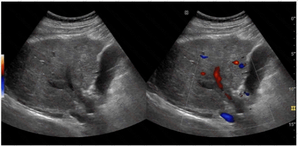
Which condition is demonstrated in this image?
A.
Cavernous transformation
B.
Portal vein thrombosis
C.
Portal hypertension
D.
Tumor extension
Full Access
Answer:
A
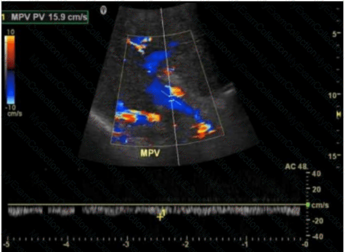
Explanation:
The image shows a color Doppler ultrasound of the main portal vein (MPV), which appears irregular and replaced by multiple small, serpiginous vascular channels — a hallmark of cavernous transformation. Cavernous transformation of the portal vein is a late complication of chronic portal vein thrombosis, in which collateral vessels develop around the thrombosed portal vein to bypass the obstruction.
Key Doppler ultrasound features of cavernous transformation:
Absence of a normal portal vein
Multiple tortuous vessels in the porta hepatis
Color Doppler shows hepatopetal flow in these channels
Low velocity, continuous waveform flow in collateral vessels
Differentiation from other options:
B. Portal vein thrombosis: Would show an absence of color flow within the portal vein lumen and possibly echogenic material within the vessel. There would be no serpiginous collateral vessels yet if it's an acute process.
C. Portal hypertension: Often diagnosed with other sonographic findings (e.g., splenomegaly, reversed portal flow, varices) but not characterized by the replacement of the portal vein by collateral vessels.
D. Tumor extension: Typically appears as echogenic intraluminal material within the portal vein with arterial waveforms on Doppler due to neovascularity. Tumor thrombus can be seen in hepatocellular carcinoma or pancreatic cancer, not multiple small collateral vessels.
[References:, Rumack CM, Wilson SR, Charboneau JW, Levine D. Diagnostic Ultrasound. 5th Edition. Elsevier, 2018. Chapter: Portal Venous System, pp. 107–110., American Institute of Ultrasound in Medicine (AIUM). Practice Parameter for the Performance of a Vascular Ultrasound Examination, 2021., Radiopaedia.org. Cavernous transformation of the portal vein: https://radiopaedia.org/articles/cavernous-transformation-of-the-portal-vein, , , ]
Question # 46
Which technique would best eliminate the spectral Doppler artifact in this image?
A.
Adjust gain
B.
Adjust baseline
C.
Increase wall filter
D.
Increase pulse repetition frequency
Full Access
Answer:
A
Explanation:
The spectral Doppler image demonstrates excessive noise along the baseline, including a “fuzzy†or filled-in spectral window. This artifact is known as spectral broadening or “blossoming,†and it typically results from excessive Doppler gain.
When Doppler gain is set too high, it amplifies not only the true Doppler signal but also the background noise. This results in a falsely broadened waveform that can obscure diagnostic information such as peak velocities or flow turbulence. The best way to correct this artifact is to reduce the Doppler gain (Option A).
Key points regarding gain-related artifact:
Excessive gain exaggerates spectral display by amplifying weak signals and noise.
Reducing gain restores the clarity of the spectral window and accurate envelope definition.
The goal is to optimize gain just enough to see the real flow signals without cluttering the display.
Differentiation from other options:
B. Adjust baseline: Useful in avoiding aliasing but does not affect gain-related noise.
C. Increase wall filter: Removes low-frequency signals from vessel wall motion but not background spectral noise.
D. Increase pulse repetition frequency (PRF): Used to reduce aliasing in high-velocity flow, not to address gain-related spectral clutter.
[References:, Kremkau FW. Sonography: Principles and Instruments. 9th Edition. Elsevier, 2015. Chapter: Doppler Principles, pp. 189–193., Rumack CM, Wilson SR, Charboneau JW, Levine D. Diagnostic Ultrasound. 5th Edition. Elsevier, 2018. Chapter: Doppler Artifacts, pp. 65–67., American Institute of Ultrasound in Medicine (AIUM) Doppler Ultrasound Practice Guidelines, 2020., —, , ]
Question # 47
Which vessel is most likely to display hepatofugal flow in the presence of portal hypertension?
A.
Coronary vein
B.
Splenic vein
C.
Inferior vena cava
D.
Inferior epigastric vein
Full Access
Answer:
A
Explanation:
The coronary vein (left gastric vein) is a common collateral pathway in portal hypertension. It often becomes dilated and may demonstrate hepatofugal (reversed) flow as blood diverts from the high-pressure portal system into systemic collaterals.
According to Zwiebel’s Introduction to Vascular Ultrasound:
“The left gastric (coronary) vein is a frequent site of hepatofugal flow in portal hypertension, reflecting collateral development.â€
[Reference:, Zwiebel WJ, Pellerito JS. Introduction to Vascular Ultrasound. 6th ed. Elsevier, 2019., AIUM Practice Parameter for Portal Venous Doppler Ultrasound, 2020.]
Question # 48
Which imaging technique best demonstrates ureteral patency?
A.
Spectral Doppler
B.
Gray scale
C.
Color Doppler
D.
Graded compression
Full Access
Answer:
C
Explanation:
Color Doppler imaging can detect ureteral jets, which represent urine entering the bladder from the ureters. The presence of bilateral ureteral jets confirms ureteral patency. Gray scale may show hydronephrosis but does not directly assess flow.
According to Rumack’s Diagnostic Ultrasound:
“Color Doppler demonstrates ureteral jets within the bladder, confirming ureteral patency.â€
[Reference:, Rumack CM, Wilson SR, Charboneau JW, Levine D. Diagnostic Ultrasound. 5th ed. Elsevier, 2017., AIUM Practice Parameter for Renal Ultrasound, 2020., —]
Question # 49
Which vessel is indicated by the arrow on this image?
A.
Proper hepatic artery
B.
Superior mesenteric artery
C.
Left renal vein
D.
Right renal artery
Full Access
Answer:
B
Explanation:
The ultrasound image demonstrates a transverse view of the abdominal vasculature, where the arrow is pointing to a circular vascular structure anterior to the aorta and posterior to the body of the pancreas — consistent with the superior mesenteric artery (SMA).
The SMA originates from the anterior aspect of the abdominal aorta just below the level of the celiac trunk and courses anterior to the left renal vein and uncinate process of the pancreas. On transverse ultrasound, it is often seen in cross-section as a round, pulsatile structure with echogenic walls, situated just anterior to the aorta. This appearance is known as the “target sign†or “bull's-eye†appearance.
Vessel Position Landmarks (transverse plane):
Aorta: Posterior and central
SMA: Just anterior to the aorta
Left renal vein: Passes between the aorta and SMA (nutcracker location)
Right renal artery: Courses posterior to the IVC toward the right kidney
Differentiation from other options:
A. Proper hepatic artery: Typically visualized within the liver hilum (portal triad), not in this anatomic location.
C. Left renal vein: Seen in transverse as a longer, oval structure crossing anterior to the aorta and posterior to the SMA.
D. Right renal artery: Arises laterally from the aorta and courses posterior to the IVC — not visualized in this axial midline location.
[References:, Rumack CM, Wilson SR, Charboneau JW, Levine D. Diagnostic Ultrasound. 5th Edition. Elsevier, 2018. Chapter: Vascular Anatomy and Abdominal Vessels, pp. 471–475., American Institute of Ultrasound in Medicine (AIUM) Practice Parameter for the Performance of an Ultrasound Examination of the Abdomen and/or Retroperitoneum, 2020., Radiopaedia.org. Superior mesenteric artery: https://radiopaedia.org/articles/superior-mesenteric-artery, —, ]